- The submandibular triangle:
- Is a subsection of the larger anterior triangle of the neck:
- Which is defined by the following boundaries:
- Lateral:
- Sternocleidomastoid muscle
- Superior:
- Inferior border of the mandible
- Medial:
- Anterior midline of the neck
- Lateral:
- Which is defined by the following boundaries:
- Is a subsection of the larger anterior triangle of the neck:
- The submandibular triangle, also known as digastric triangle:
- Is located superior to the hyoid bone
- It is bordered:
- Superiorly by the inferior border of the mandible and the mastoid process
- Posteriorly by the posterior belly of the diagastric and stylohoid muscles
- Anteriorly by the anterior belly of digastric muscle
- The roof of the triangle is formed by the:
- Skin
- Superficial cervical fascia
- The platysma
- Deep cervical fascia
- The branches of the facial nerve and transverse cutaneous cervical nerves:
- Also pass over the roof of the triangle
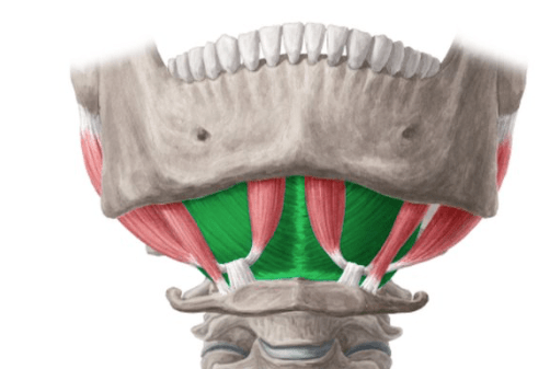
- Digastric muscle:
- The submandibular triangle is largely defined by the digastric muscle:
- Which is a double-bellied muscle that depresses the mandible:
- Opens the mouth
- Which is a double-bellied muscle that depresses the mandible:
- The anterior belly:
- Arises from the digastric fossa found in the inner / internal aspect of the anterior mandible
- The posterior belly:
- Arises from the mastoid notch of temporal bone
- Both are joined by a tendinous sheath:
- Attach to the hyoid bone
- The submandibular triangle is largely defined by the digastric muscle:

- A major landmark of the submandibular triangle:
- Is the submandibular gland (innervated by the facial nerve):
- This salivary gland can be described as having two lobes:
- Which are divided by the posterior border of the mylohyoid muscle
- The superificial lobe:
- Is the larger of the two
- Located superficial to the inferior surface of the mylohyoid muscle
- The smaller deep lobe wraps around the posterior border of the mylohyoid
- This salivary gland can be described as having two lobes:
- Is the submandibular gland (innervated by the facial nerve):
- Contents of the submandibular triangle:
- In terms of surgical practice, the submandibular triangle is best visualized as having four layers:
- These layers start from the skin and continue progressively deeper
- First layer (roof):
- As previously mentioned, the roof of the submandibular triangle i.e. the first plane encountered surgically comprises of the skin and the superficial fascia:
- These enclose the platysma muscle and the subcutaneous fat
- Also enclosed are the cervical and mandibular branches of the facial nerve (cranial nerve VII)
- As previously mentioned, the roof of the submandibular triangle i.e. the first plane encountered surgically comprises of the skin and the superficial fascia:
- Second layer (submandubilar space):
- The second surgical plane of the submandibular triangle, the following contents can be found:
- The submandibular lymph nodes
- The superficial portion / lobe of the submandibular gland
- The submental branch of the facial vein:
- Which accompanies the submental branch of the facial artery
- The vessels and nerves to mylohyoid muscle:
- Lie directly along the inferior surface of the same muscle
- The superficial / investing layer of the deep cervical fascia is also located here
- Both the facial vein and anterior branch of the retromandibular vein:
- Cross the triangle anterior, or superficial to the submandibular gland, and unite near to the angle of the mandible:
- To form the common facial vein:
- The common facial vein then drains into the internal jugular vein near the greater cornu of the hyoid bone
- To form the common facial vein:
- Cross the triangle anterior, or superficial to the submandibular gland, and unite near to the angle of the mandible:
- The facial artery (which is the fourth branch of the external carotid artery):
- Also enters the submandibular triangle by passing beneath the posterior belly of the digastric muscle, as well as the stylohyoid muscle
- Once it enters the triangle, it also lies deep to the submandibular gland
- Once the artery has crossed the gland over its posterior aspect, it curls around the inferior border of the mandible, and ascends superomedially across the facial region
- The inferior tip of the parotid gland can be found within in the posterior region of the digastric triangle
- Ascending within the substance of the parotid gland is the external carotid artery
- The second surgical plane of the submandibular triangle, the following contents can be found:
- Third layer (floor):
- Next is the third surgical layer
- Once again the structures from superficial to deep are the:
- Mylohyoid muscle along with its nerve
- The hyoglossus muscle
- As well as the middle pharyngeal constrictor muscle:
- Which lies over the lower part of the superior pharyngeal constrictor, and a subsection of the styloglossus muscle
- The mylohyoid muscles:
- Are regarded as the true diaphragm of the floor of the mouth
- These muscles arise from the mylohyoid line:
- That is found on the inner surface of the mandible
- Inserts into the body of the hyoid bone itself
- The nerve that supplies the mylohyoid:
- Is a branch of the alveolar division of the mandibular division of the trigeminal nerve (CN V3):
- Lies on the surface of the inferior aspect of the muscle
- Is a branch of the alveolar division of the mandibular division of the trigeminal nerve (CN V3):
- The superior surface of mylohyoid is in contact with the lingual nerve (division of V3) and hypoglossal nerve (cranial nerve XII)
- In terms of surgical practice, the submandibular triangle is best visualized as having four layers:
- Fourth layer (basement / sublingual space)
- Finally we have the deepest or fourth surgical plane
- The structures within this plane, from superficial to deep are:
- The deep portion of the submandibular gland
- The duct of the submandibular gland (Wharton’s duct)
- The lingual nerve (division of V3)
- The sublingual artery & vein:
- Which lie superficial to the sublingual gland
- The submandibular duct is found inferior to the lingual nerve (except where the lingual nerve passes beneath it) as well as superior to the hypoglossal nerve
- Deeper still we find cranial nerve XII (hypoglossal nerve), as well as the submandibular ganglion

