👉Ductal calcifications have a wide variety of presentations depending upon the underlying process that created them.
👉When coarse rod-like ductal calcifications are diffuse, bilateral, and not confined to a single lobe, they can be confidently assumed to result from plasma cell mastitis, and they do not require further evaluation or biopsy.
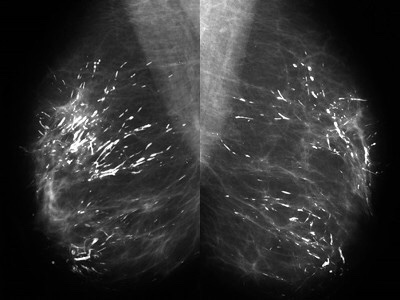
👉The process is called secretory disease because there is a stagnant, viscous fluid that eventually petrifies and results in the smooth contoured calcifications.
👉Some of them are branching and look like malignant casting type calcifications, but the key distinguishing feature is the diffuse, multilobe, bilateral nature of the process.
👉Calcifications become much more worrisome when they are confined to a single lobe.
👉The most frequent malignant, ductal “casting type” calcifications are fragmented, linear, and branching, and they are the most reliable mammographic sign of malignancy.
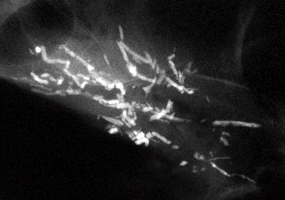
👉The presence of fragmented and/or dotted casting type calcifications on the mammogram restricted to one lobe is a pathognomonic sign of a diffuse, grade 3 breast cancer subtype that originates in the major ducts and usually has a solid or micropapillary pattern.
👉Traditionally, this subtype has been called “comedo carcinoma.”
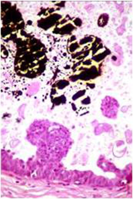
👉The cancer cells either produce a viscous, proteinaceous fluid which gradually concentrates and eventually calcifies, or they undergo necrosis (apoptosis) followed by calcification.
👉In both instances the intraluminal pressure increases, distending the ducts considerably.
👉Dotted casting type calcifications have been referred to as “snake skin-like calcifications” and they accumulate in the fluid produced by either micropapillary or solid cancer cell growth patterns.
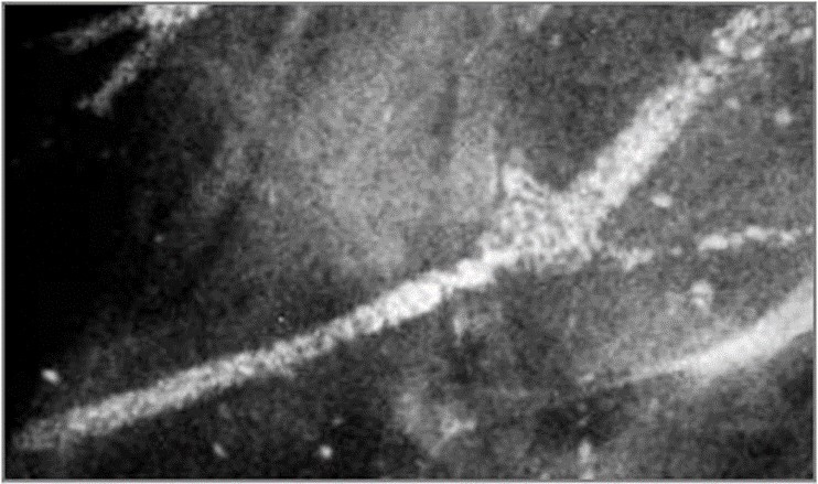
👉The tips of the micropapillary growths may become detached and eventually calcify, contributing to the intraluminal calcifications.

 Rodrigo Arrangoiz MS, MD, FACS cirujano oncology y cirujano de mamá de Sociedad Quirúrgica S.C en el America British Cowdray Medical Center en la ciudad de Mexico:
-
Es experto en el manejo del cáncer de mama.

Es miembro de la American Society of Breast Surgeons:

Training:
• General surgery:
• Michigan State University:
• 2004 al 2010

• Surgical Oncology / Head and Neck Surgery / Endocrine Surgery:
• Fox Chase Cancer Center (Filadelfia):
• 2010 al 2012

• Masters in Science (Clinical research for health professionals):
• Drexel University (Filadelfia):
• 2010 al 2012

• Surgical Oncology / Head and Neck Surgery / Endocrine Surgery:
• IFHNOS / Memorial Sloan Kettering Cancer Center:
• 2014 al 2016