-
Overview:
-
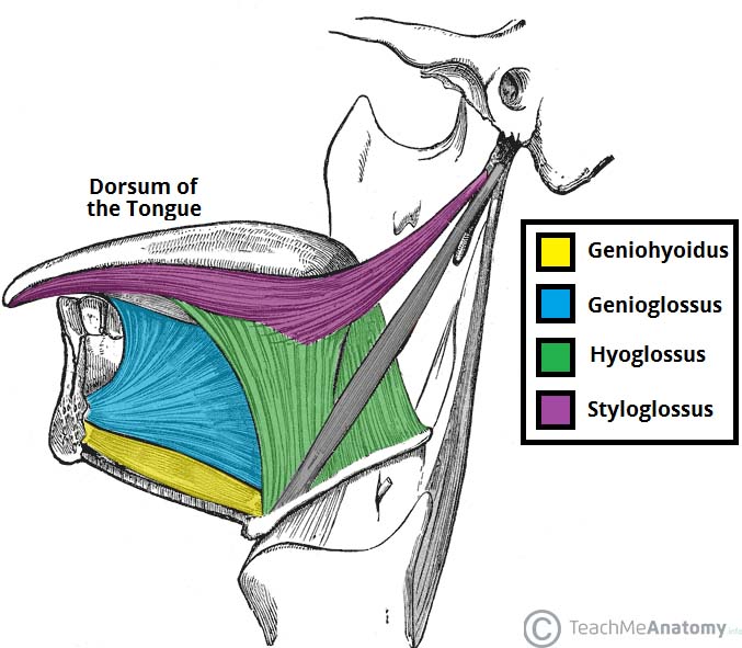
The tongue is a mass of muscle that is almost completely covered by a mucous membrane.
-
It occupies most of the oral cavity and oropharynx.
-
It is known for its role in taste, but it also assists with mastication (chewing), deglutition (swallowing), articulation (speech), and oral cleansing.
-
Five cranial nerves contribute to the complex innervation of this multifunctional organ.
-
The embryologic origins of the tongue first appear at 4 weeks’ gestation:
-
The body of the tongue forms from derivatives of the first branchial arch:
-
This gives rise to two lateral lingual swellings and one median swelling (known as the tuberculum impar):
-
The lateral lingual swellings slowly grow over the tuberculum impar and merge, forming the anterior two thirds of the tongue.
-
-
-
Parts of the second, third, and fourth branchial arches give rise to the base of the tongue.
-
-
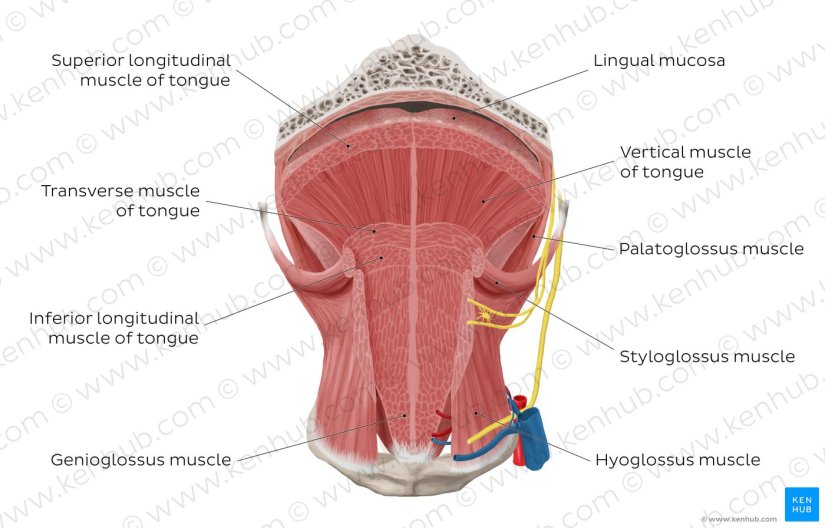
Occipital somites give rise to myoblasts, which form the intrinsic tongue musculature.
-
-
Gross Anatomy:
-
From anterior to posterior, the tongue has 3 surfaces:
-
Tip
-
Body
-
Base.
-
-
The tip is the highly mobile, pointed anterior portion of the tongue.
-
Posterior to the tip lies the body of the tongue, which has dorsal (superior) and ventral (inferior) surfaces.
-
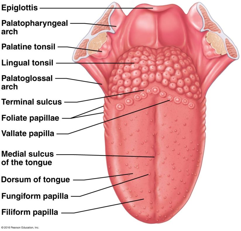
The median sulcus of the tongue separates the body into left and right halves.
-
The terminal sulcus, or groove, is a V-shaped furrow that separates the body from the base of the tongue:
-
At the tip of this sulcus is the foramen cecum, a remnant of the proximal thyroglossal duct.
-
-
The base of tongue contains the lingual tonsils, the inferiormost portion of Waldeyer’s ring.
-
-
Lingual papillae:
-
The surface of the body of the tongue derives its characteristic appearance from the presence of lingual papillae:
-
Which are projections of lamina propria covered with epithelium.
-
-
The four types of lingual papillae are as follows:
-
Vallate (circumvallate)
-
Foliate
-
Filiform
-
Fungiform
-
-
The vallate papillae (circumvallate) are flat, prominent papillae that are surrounded by troughs:
-
In humans, there are 8 to 12 vallate papillae, located directly anterior to the terminal sulcus.
-
The ducts of the lingual glands of von Ebner secrete lingual lipase into the surrounding troughs to begin the process of lipolysis.
-
-
-
The foliate papillae are small folds of mucosa (short vertical folds) located along the lateral surface of the tongue:
-
They are located on the sides at the back of the tongue, just in front of the palatoglossal arch of the fauces:
-
The foliate papillae appear as a series of red colored, leaf–like ridges of mucosa.
-
-
There are four or five vertical folds, and their size and shape is variable.
-
They are covered with epithelium, lack keratin and so are softer, and bear many taste buds:
-
Approximately 1000 taste buds.
-
Taste buds, the receptors of the gustatory sense, are scattered over the mucous membrane of their surface.
-
-
-
They are usually bilaterally symmetrical.
-
Sometimes they appear small and inconspicuous, and at other times they are prominent.
-
Because their location is a high risk site for oral cancer, and their tendency to occasionally swell, they may be mistaken as tumors or inflammatory disease.
-
Serous glands drain into the folds and clean the taste buds.
-
Lingual tonsils are found immediately behind the foliate papillae and, when hyperplastic, cause a prominence of the papillae.
-
-
The filiform papillae:
-
Are the most numerous of the lingual papillae.
-
They are fine, small, cone-shaped papillae covering most of the dorsum of the tongue.
-
They cover most of the front two-thirds of the tongue’s surface.
-
They appear as very small, conical or cylindrical surface projections, and are arranged in rows which lie parallel to the sulcus terminalis:
-
At the tip of the tongue, these rows become more transverse.
-
-
They are responsible for giving the tongue its texture and are responsible for the sensation of touch.
-
Unlike the other kinds of papillae, filiform papillae do not contain taste buds.
-
-
The fungiform papillae are mushroom shaped (generally red in color) and are dispersed most densely along the tip and lateral surfaces of the tongue:
-
Humans have approximately 200 to 300 fungiform papillae.
-
-
Each vallate, foliate, and fungiform papilla contains taste buds (250, 1000, and 1600 taste buds, respectively):
-
Each taste bud is innervated by several nerve fibers.
-
In humans, all taste buds can perceive the five different taste qualities:
-
Salt
-
Sweet
-
Bitter
-
Acid
-
Umami.
-
-
-
-
Each taste bud consists of taste receptor, basal cell , and edge cells.
-
When a taste molecule binds to a taste receptor, the receptor cell depolarizes:
-
Causing an influx of Ca++, which results in the release of an unknown neurotransmitter.
-
Following depolarization, the afferent neural pathway depends on the location of the taste bud that was stimulated:
-
In the anterior two thirds of the tongue, the chorda tympani branch of the facial nerve (cranial nerve VII) is stimulated.
-
The lingual-tonsillar branch of the glossopharyngeal nerve (cranial nerve IX) relays taste information from the posterior third of the tongue.
-
-
-
Taste fibers from the anterior two thirds of the tongue first travel with the lingual nerve and then are relayed to the chorda tympani nerve:
-
This nerve enters the temporal bone from the infratemporal fossa, where it joins the facial nerve and travels to the geniculate ganglion, where its pseudounipolar cell bodies are located.
-
From the geniculate ganglion the taste fibers travel in the nervus intermedius to the nucleus of the solitary tract located in the medulla oblongata.
-
-
Similarly, taste fibers from the posterior one third of the tongue travel with the lingual-tonsillar nerve to the inferior glossopharyngeal ganglion and then to the nucleus of the solitary tract located in the medulla oblongata.
-
Second-order neurons then project taste fibers to the parabrachial nucleus of the pons.
-
The central tegmental tract carries taste sensation from the pons to the thalamus.
-
The pathway ends in the frontal operculum and insular cortex.


Rodrigo Arrangoiz MS, MD, FACS a head and neck surgeon and is amember of Sociedad Quirúrgica S.C at the America British Cowdray Medical Center.
He is first author on some publications on oral cavity cancer:
-
Oral Tongue Cancer: Literature Review and Current Management
-
Understand Cancer: Research and Treatment Oral Cavity Cancer: Literature Review and Current Management.


